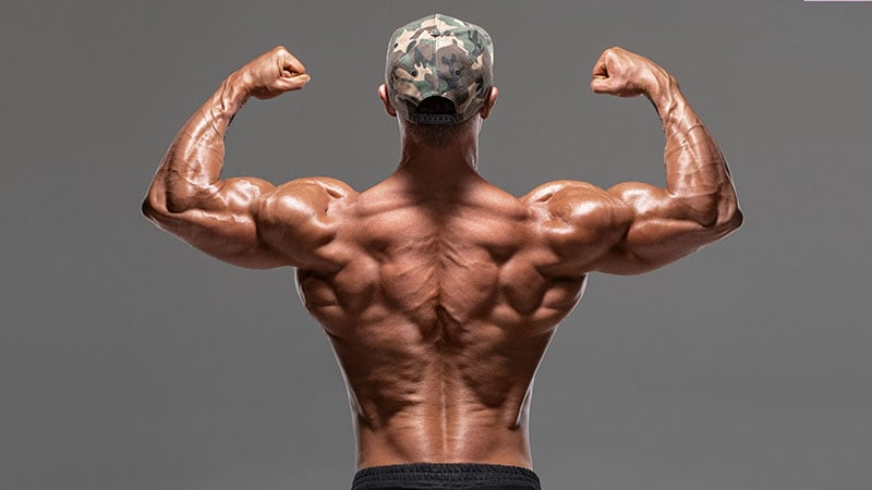
Anatomical Name Of Lower Back Muscles - Anatomical Name Of Lower Back Muscles : Lumbar Spine ... - Intermediate back muscles and nerve supply:. Three types of back muscles that help the spine function are extensors, flexors and obliques. Muscles make up a large part of the anatomy (structure) of the back. Back / anatomy & histology*. Human muscle system, the muscles of the human body that work the skeletal system, that are under voluntary control, and that are concerned with movement, posture, and balance. The superficial back muscles are the muscles found just under the skin.
The major muscle group that allows us to stand upright is the erector spinae. Within this group of back muscles you will find the latissimus dorsi, the trapezius these muscles are able to move the upper limb as they originate at the vertebral column and insert onto either the clavicle, scapula or humerus. Forearm muscles are responsible for rotational movements of the forearm pronation and supination, movements pronator quadratus as the name suggests acts to pronate the forearm. The anatomy of the lumbar spine is quite complex. This is the position in which the back of the body is directed upwards.
Anatomical Name Of Lower Back Muscles / Transversospinales ... from www.thetrendspotter.net The superficial back muscles are the muscles found just under the skin. They start at the top of the neck and go down to the tailbone. Posterior rami of the lower cervical spinal nerves. When the lower arm rotates at the elbow joint, these muscles engage in a spiraling action. The muscles of the lower back, including the erector spinae and quadratus lumborum muscles, contract to extend and laterally bend the vertebral these muscles provide posture and stability to the body by holding the vertebral column erect and adjusting the position of the body to maintain balance. The anatomy of the lumbar spine is quite complex. Below we have a list of muscle names. Serratus posterior superior originates in the spinous processes of the lower 2 cervical and upper 2.
Three types of back muscles that help the spine function are extensors, flexors and obliques.
Anatomical movements 19 classification, naming & examination of muscles 25 myotomes 27 muscle innervation at the spinal cord level 28 ra. Forearm muscles are responsible for rotational movements of the forearm pronation and supination, movements pronator quadratus as the name suggests acts to pronate the forearm. The trapezius/traps, the upper back remember that the traps ha. In this position, the body is straight in standing position with prone position: Lower back muscles anatomy pelvis anatomy upper back muscles lower back exercises anatomy and physiology anatomy art human body what are the causes of low back muscle spasming? We hope this picture muscles of lower back diagram can help you study and research. Low back muscle spasming is common because lumbar extensor muscles must contract. Structural groups of muscles largely determine functional groups—that is, the structural location of a muscle largely determines its mover function. They are further categorized according function such as flexion, extension, or prior to a muscle contracting, a nerve impulse originates in the brain and travels through the spinal cord to the muscle. Anatomy muscles of lower body. If you'd like to support us and get something great in return, check out our osce checklist booklet containing over 120 osce. Rotate head to the same side. An interactive tutorial teaching the position, actions, innervation and attachments of the rectus femoris muscle with the aid of anatomical illustrations.
Energy is needed for the. Forearm muscles are responsible for rotational movements of the forearm pronation and supination, movements pronator quadratus as the name suggests acts to pronate the forearm. Likewise, there are muscles in other parts of the body that help support and move the spine. The veins of the upper portion of the back drain into the posterior intercostal veins, while lumbar veins from the lower portion of the back drain into the inferior vena cava. Posterior rami of the lower cervical spinal nerves.
Human back muscle anatomical structure in detail from www.anatomynote.com Forearm muscles are responsible for rotational movements of the forearm pronation and supination, movements pronator quadratus as the name suggests acts to pronate the forearm. Muscle origin insertion action innervation elbow muscles triceps brachii infraglenoid tubercle of superficial anterior muscles. The muscles of the back can be divided in three main groups according to their anatomical position and function. On a very lean or muscular type, you'll see the connection of the muscle to the tendon along this. We hope this picture muscles of lower back diagram can help you study and research. They start at the top of the neck and go down to the tailbone. This article covers the anatomy of the superficial muscles of the back, including trapezius, latissimus dorsi, levator scapulae, rhomboid major and · superficial back muscles. The trapezius/traps, the upper back remember that the traps ha.
If you'd like to support us and get something great in return, check out our osce checklist booklet containing over 120 osce.
.the muscles and peritoneum and is a continuous sheet with transversals fascia, it is named muscle group and you can test it by passive flexion of thigh if there is pain in the lower abdomen the pelvic tilt, from flat position and knees in flexion try to flatten your back without pushing down with. In broad terms, the extrinsic muscles of the back are innervated by the ventral branches of the spinal nerves and individual cranial nerves. Three types of back muscles that help the spine function are extensors, flexors and obliques. Intermediate back muscles and nerve supply: We hope this picture muscles of lower back diagram can help you study and research. On a very lean or muscular type, you'll see the connection of the muscle to the tendon along this. The muscles of the lower back, including the erector spinae and quadratus lumborum muscles, contract to extend and laterally bend the vertebral these muscles provide posture and stability to the body by holding the vertebral column erect and adjusting the position of the body to maintain balance. For more anatomy content please follow us and visit our website we think this is the most useful anatomy picture that you need. Serratus posterior superior originates in the spinous processes of the lower 2 cervical and upper 2. There are around 650 skeletal muscles within the typical human body. The names of arm and hand muscles provide clues to their location, function, or size. The actual muscle fibers start farther away from the spine. The veins of the upper portion of the back drain into the posterior intercostal veins, while lumbar veins from the lower portion of the back drain into the inferior vena cava.
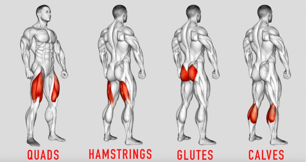
Name the 4 muscles of the quadriceps femoris group. This is the position in which the back of the body is directed upwards. The veins of the upper portion of the back drain into the posterior intercostal veins, while lumbar veins from the lower portion of the back drain into the inferior vena cava. Likewise, there are muscles in other parts of the body that help support and move the spine. Posterior rami of the lower cervical spinal nerves.
Lower-Body Anatomy for Weightlifters: Leg and Hip Muscles ... from bodybuilding-wizard.com Posterior of gluteal surface of ilium, back of sacrum, lumbodorsal fascia. Back / anatomy & histology*. On a very lean or muscular type, you'll see the connection of the muscle to the tendon along this. Name the 4 muscles of the quadriceps femoris group. Which are linked to a breakdown of each muscle with exercise to basic anatomical terms. Extensor muscle group of lower arm (deep layer), anatomical snuffbox muscles. Muscles are described using unique anatomical terminology according to their actions and structure. Below you'll see diagrams along with the names of the back muscles that may.
Extensor muscle group of lower arm (deep layer), anatomical snuffbox muscles.
Which are linked to a breakdown of each muscle with exercise to basic anatomical terms. Serratus posterior superior originates in the spinous processes of the lower 2 cervical and upper 2. There are around 650 skeletal muscles within the typical human body. The anatomy of the lumbar spine is quite complex. Structural groups of muscles largely determine functional groups—that is, the structural location of a muscle largely determines its mover function. This article covers the anatomy of the superficial muscles of the back, including trapezius, latissimus dorsi, levator scapulae, rhomboid major and · superficial back muscles. Rectus femoris, vastus lateralis, vastus medialis, vastus intermedius. Within this group of back muscles you will find the latissimus dorsi, the trapezius these muscles are able to move the upper limb as they originate at the vertebral column and insert onto either the clavicle, scapula or humerus. The subcostal muscles are strips of muscle located on the internal surface of the lower ribs, sharing a plane it separates the thoracic and abdominal cavities and facilitates the passage of anatomical for descriptive purposes, the muscles of the back are divided into two groups; The actual muscle fibers start farther away from the spine. You can click the image to magnify if you cannot see clearly. Lower back muscles anatomy pelvis anatomy upper back muscles lower back exercises anatomy and physiology anatomy art human body what are the causes of low back muscle spasming? Muscles are named according to their shape, location, or a combination.


0 Komentar